
90 years of vectorcardiography (VKG)
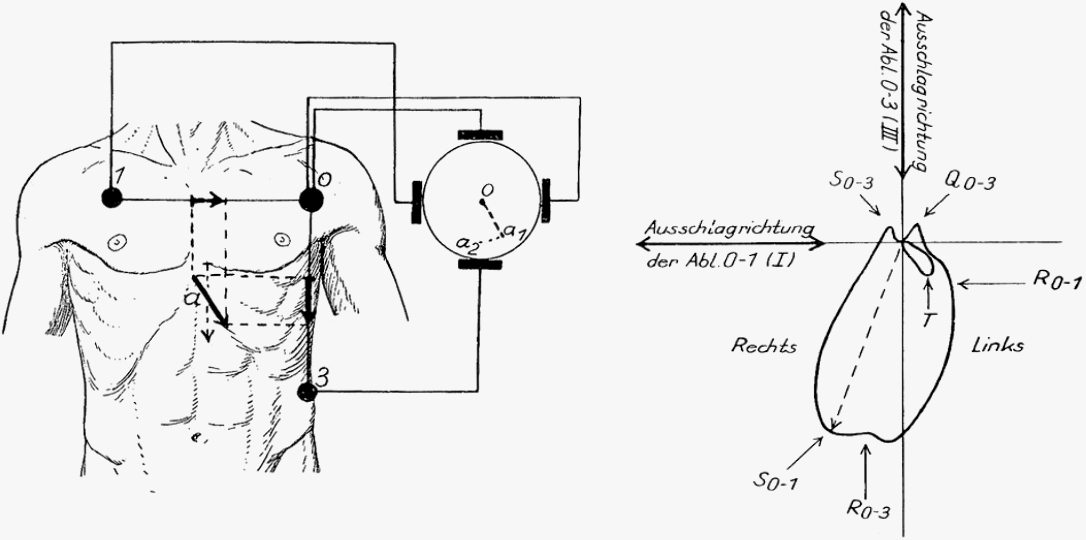
The introduction of vectorcardiography (VCG) by the German internist and cardiologist Fritz Schellong in 1936 marked a milestone in non-invasive cardiac diagnostics. A pioneer of clinical research on circulatory regulation and electrocardiography, Schellong developed a dedicated apparatus and a novel lead system to capture the spatial propagation of cardiac electrical excitation.
Rather than recording only the temporal voltage changes as in conventional electrocardiography (ECG), Schellong enabled the visualization of vector loops in the frontal plane. He demonstrated the method in healthy individuals as well as in cases of left axis deviation and left ventricular hypertrophy.
By integrating contemporary advances in physics to complement electrocardiography, Schellong showed that VCG represents a valuable method for the analysis of disorders of circulatory function. Fritz Schellong (1891–1953) is therefore regarded as one of the pioneers of clinical vectorcardiography.
Today, vectorcardiography is experiencing a renaissance through its integration with artificial intelligence. Cardisiography represents a modern advancement that enables three-dimensional analysis of cardiac activity using a limited number of electrodes. AI-based algorithms facilitate faster and earlier detection of cardiac pathologies, such as coronary heart disease.
Advantages: Compared with exercise ECG, Cardisiography demonstrates high sensitivity (>90%) and enables precise diagnosis of ischemia-related disturbances of myocardial repolarization. Vectorcardiography is thus becoming established as a valuable non-invasive method for advanced cardiac diagnostics.
Learn more about Cardisiography: https://cardis.io/en/application/cardisiography/