Cardisiography (CSG) Detects
Using artificial intelligence and the scientific concept of vectorcardiography, our technology detects:
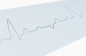
Rhytmological Diseases
Cardiac arrhythmias (rhythmology) are noticed by patients as an abnormally beating heart. Physicians can distinguish between the slow heartbeat (bradycardia), the misguided heartbeat (bundle branch block) and the fast heartbeat (tachycardia).
Coronary Diseases
In coronary heart disease (CHD), the coronary arteries are narrowed and calcified as a result of atherosclerosis. Atherosclerosis refers to the pathological storage of cholesterol esters and other fats in the inner wall layer of arterial blood vessels. This inflammatory process can begin at a young age.
Structural Diseases
“Structural heart disease” refers to all diseases of the heart involving a physical defect in the valves or the walls of the heart, affecting it’s functioning.
Pathological Patterns
Developed as a quick, precise, and non-invasive tool, Cardisiography (CSG) detects over 30 pathological electrical patterns, indicating heart disease.
Rhythms
Sinus Bradycardia
Sinus Rhythm
Sinus Tachycardia
Pacemaker Rhythm
Atrial Fibrillation
Atrial Fibrillation with Rapid Ventricular Response
Atrial Fibrillation with Slow Ventricular Response
Atrial Flutter
Atrial Flutter with Rapid Ventricular Response
Atrial Flutter with Slow Ventricular Response
Supraventricular Tachycardia
Suspected Junctional Rhythm
Suspected Junctional Bradycardia
Suspected Accelerated Junctional Rhythm
Wide QRS Rhythm
Idioventricular Rhythm
Wide QRS Tachycardia
Conduction Anomalies
First-Degree AV Block
Second-Degree AV Block, Wenckebach Type
Higher-Degree AV Block
Complete Right Bundle Branch Block
Incomplete Right Bundle Branch Block
Complete Left Bundle Branch Block
Incomplete Left Bundle Branch Block
Nonspecific Intraventricular Conduction Delay
Left Anterior Fascicular Block
Left Posterior Fascicular Block
Bifascicular Block (RBBB + LAFB)
Bifascicular Block (RBBB + LPFB)
Trifascicular Block (RBBB + LAFB + AV Block 1)
Trifascicular Block (RBBB + LPFB + AV Block 1)
Ectopy
Premature Complexes
Axes
Left Axis Deviation
Right Axis Deviation
Extreme Axis Deviation
Normal Axis
Infarctions
Suspected ST-Elevation ACS (Acute Coronary Syndrome)
Suspected Non-ST-Elevation ACS (Acute Coronary Syndrome)
Occlusion Myocardial Infarction (OMI) – Available as Plan Plus (OMI) Only
Hypertrophies
Suspected Atrial Enlargement
Suspected Ventricular Hypertrophy
Other
Suspected Long QT Syndrome
Suspected Short QT Syndrome
Causes & Symptoms
Reduced blood flow and agitation disorders of the heart can have various causes. Early detection of the underlying disease is crucial for a good prognosis. This information is general advice and must not be used for self-diagnosis or self-treatment and cannot replace a visit to the doctor.
Cardiac arrhythmias (arrhythmia)
Find out more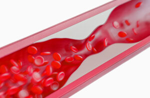
Stenosis
Find out more
Heart valve defects
Find out more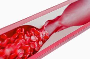
Angina pectoris
Find out more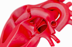
Coronary heart disease (CHD)
Find out more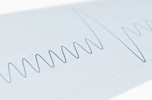
Shortness of breath, heart stumbling and dizziness
Find out more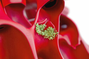
Inflammation of the heart muscle or valvular apparatus
Find out more