
Resultados respaldados por estudios científicos
La cardiosiografía destaca por su capacidad única para detectar enfermedades cardíacas en fases muy tempranas, incluso en personas completamente asintomáticas. En casos donde el estado general de salud no permite evaluar con precisión el flujo sanguíneo al
corazón, este método ofrece una solución innovadora y eficaz.
Diversos estudios han demostrado de forma contundente los beneficios de la cardiosiografía para identificar precozmente la disminución del riego sanguíneo al músculo cardíaco, lo que permite actuar antes de que se presenten complicaciones graves.
A continuación, puedes consultar un resumen detallado con los fundamentos científicos y los principales hallazgos de nuestra investigación:
Cardisio en PubMed
![]()
Actual e importante
New VCG Algorithms for the Detection of LV Hypertrophy and Impaired LV Function
hhh
Evaluation of new vectorcardiography algorithms for identifying left ventricular hypertrophy and impaired systolic function
Janek Salatzki, Arne Kristian Schwarz, Sarah Wolfsteller, Nicolas Hein, Erika Poyo Medina, Norbert Frey, Henning Steen, Florian André, Alexander Passow. Universitätsklinikum Heidelberg, Journal of Electrocardiology, Volume 97, July–August 2026, 154272
1. Background and Clinical Relevance
Heart failure with reduced ejection fraction (HFrEF) and left ventricular hypertrophy (LVH) represent two of the most prevalent and prognostically significant forms of cardiac structural remodeling. Despite their substantial clinical impact, both conditions are frequently diagnosed at an advanced stage, partly because currently available screening modalities are either resource-intensive (echocardiography, cardiac magnetic resonance imaging [CMR]) or diagnostically limited (conventional 12-lead electrocardiography).
Conventional electrocardiography provides only limited sensitivity for detecting subtle structural cardiac abnormalities due to its inherently two-dimensional representation of cardiac electrical activity. In contrast, vectorcardiography (VCG) captures cardiac electrical activity in three orthogonal planes, enabling a substantially more comprehensive spatiotemporal assessment of depolarization and repolarization dynamics.
2. Study Design and Methods
Study Design and Population
This prospective case–control study was conducted at Heidelberg University Hospital between January 2023 and February 2024. A total of 245 participants undergoing both CMR and VCG examinations were included.
The study population comprised:
- Group 1 – Reduced left ventricular ejection fraction (LVEF < 40%): n = 40
- Group 2 – Left ventricular hypertrophy (indexed LV mass ≥ 55 g/m²): n = 208
- Group 3 – Combined phenotype (reduced LVEF and LVH): n = 34
- Group 4 – Control group (structurally normal hearts on CMR): n = 31
VCG Acquisition System: Cardisiography (CSG)
VCG recordings were obtained using a five-electrode Cardisiography system during a standardized 4-minute resting acquisition. An artificial intelligence (AI)-based algorithm extracted a total of 583 parameters from the raw signal data, including geometric, spatial, and temporal descriptors of depolarization and repolarization processes, such as vector magnitude, azimuth angles, conduction velocities, and loop morphology characteristics.
Reference Standard
Cardiac magnetic resonance imaging served as the reference standard. Examinations were performed on 1.5-T and 3-T Philips Ingenia scanners using standardized volumetric assessment protocols and late gadolinium enhancement (LGE) imaging for myocardial tissue characterization.
3. Results
Key Diagnostic Parameters
Among the 583 VCG-derived variables, the most discriminative parameters for structural cardiac abnormalities were identified as follows:
- Reduced LVEF: Repolarization time-difference ratio (Rpeak–Tonset / QRSend–Tpeak)
- Area under the receiver operating characteristic curve (AUC): 0.843
- Left ventricular hypertrophy: Combination of T-wave azimuth, T-wave magnitude, and azimuth variability
- AUC range: 0.739–0.791
- Combined phenotype (reduced LVEF + LVH): Repolarization time-difference ratio
- AUC: 0.866
Diagnostic Metrics at a Glance
4. Conclusions
This study provides a methodologically robust proof-of-concept for the diagnostic utility of AI-enhanced vectorcardiography in the detection of structural heart disease.
From a clinical perspective, several findings deserve particular attention:
- Strength of the technology: The method requires neither specialist interpretation nor exposure to ionizing radiation or contrast agents. A standardized 4-minute recording combined with fully automated analysis enables efficient and scalable deployment in primary care practices, emergency departments, and resource-limited healthcare settings.
- Addressing a diagnostic gap: Early identification of LVH and impaired left ventricular systolic function remains challenging with conventional ECG, particularly before the onset of clinical symptoms. AI-assisted VCG may represent a practical and scalable bridging technology for earlier detection of structural cardiac disease.
✓ Immediate PDF-based diagnostic report including an individualized risk score
✓ CE-certified medical technology compliant with GDPR requirements (DSGVO)
A mathematical approach to demonstrate R to T wave concordance of the human ECG
![]()
R-to-T-wave concordance within the same lead of the human electrocardiogram (ECG) has been under discussion for decades, as the QRS complex with its R-wave represent depolarization and the T-wave repolarization. Extracellular recorded monophasic action potential (MAP) of the human heart muscle fibre resembles the first derivation of the intracellular MAP over time, showing R-to-T-wave discordance. While a single fibre monophasic electrophysiology lacks many aspects of the ECG, bipolar registration for the different layers of the ventricular wall (transmural gradient) gives more detailed information about the local MAP, as endo-, meso- and epicardium show a MAP time difference (voltage gradient) dependent positioning of the T-wave, within a simultaneously recorded epicardial ECG.
Without an integrated consideration of the heterogenous (endo-, meso- and epimyocardial) MAP, T-wave concordance cannot be explained, as it would provide a homogenous model like the single heart muscle fibre MAP. A closed form representation of the potential difference to explain concordance was found. We developed a 3-dimensional, time dependent setup simulating the transmural gradient. The time-dependent spatial integrals, which account for the extracellular loading-density (which is inversely related to MAP) along all layers show concordance.
These functions allow to calculate the electric potential at any point in space at any given time within the QT-Interval. The closed form solution of the electric potential enables to identify the corresponding T-wave morphologies. Inversely, pathological patterns in the action potential can then be identified from the ECG. Our approach is an attempt to overcome the inverse problem and to reduce empiricism, as change of T-wave morphology can be assigned.
Community-based cardiovascular risk assessment using the CardisioTM AI test: a prospective cohort study
![]()
Background: Cardiovascular disease (CVD) accounts for significant morbidity and mortality disproportionately affecting hard-to-reach individuals. New technology that enables community testing rather than attending hospital may address health inequalities and facilitate new care pathways.
Aim: To explore whether the Cardisio test, which interprets three-dimensional vectorcardiography activity using a cloud-based artificial intelligence (AI) algorithm, can identify asymptomatic CVD.
Design & setting: Prospective cohort study in three settings: general practice, pharmacy, and a community health centre. Recruitment targeted asymptomatic adults aged ≥18 years, with a QRISK3 score ≥10% or CVD risk factors.
Method: A 10-minute test using five electrodes (four chest, one back). The Cardisio results are classified into red, amber, or green based on the Cardisio test’s perfusion (P), structure (S), and arrhythmia (A) parameters. Pre- and post-test questionnaires provided feedback on participants’ experiences. Results reviewed by a chief investigator ([CI] independent consultant cardiologist) and dealt with according to the study participants’ results and medical profile.
Results: In total, 628 tests were performed, 51% male (n = 320), 49% (n = 308) female, with a mean age of 54 years (18-75 years). In the opinion of the CI, there was a strong association between one or more Cardisio red test results and referral to cardiology clinic being indicated (P<0.001). The test was understood as easy to perform, with an 87.5% recommendation rate among participants (n = 492 of the 560).
Conclusion: This simple, near-patient test afforded high-risk hard-to-reach individuals with access to an acceptable test that can facilitate appropriate referral. The automated test does not rely on interpretation of electrocardiogram (ECG) readouts and so is more effective at identifying underlying CVD than a traditional 12-lead ECG.
Keywords: cardiovascular diseases; clinical diagnosis; health inequities; inequalities; primary health care.
Visión general
Estudio multicéntrico – Comparación de la cardisiografía (CSG) con la angiografía coronaria en pacientes con sospecha de enfermedad coronaria
![]()
Frankfurt, 16 de enero de 2020: En el número actual, la reconocida revista “Journal of Electrocardiology” publica un estudio revisado por pares que demuestra la impresionante precisión de la cardiosiografía en la detección de enfermedades coronarias. El artículo proporciona resultados sobre la sensibilidad y especificidad de la cardiosiografía: La sensibilidad es del 97% para los hombres y del 90% para las mujeres, es decir, el 97% de los hombres enfermos son reconocidos como enfermos y el 90% de las mujeres. En cuanto a la especificidad, las mujeres que participaron en el estudio superan a los hombres con un 74% y un 76%, es decir, el 76% de las mujeres sanas son reconocidas como sanas y el 74% de los hombres. La cardiografía es el primer procedimiento que se puede utilizar para determinar de forma no invasiva, rápida y económica el riesgo de enfermedad coronaria (CHD) en personas sin síntomas.
Estudio de validación de Cardiosio Sana Heart Center Cottbus
![]()
Frankfurt, 10 de marzo de 2020: Por segunda vez en poco tiempo, un estudio clínico ha llegado a la conclusión de que la cardiosiografía logra resultados comparables en la detección de enfermedades coronarias (CHD) a los del estándar de oro actual, la angiografía coronaria.
El equipo alrededor del Dr. Temirlan Erkenov del Departamento de Cirugía Cardíaca del Centro Cardíaco SANA en Cottbus, Alemania, concluyó que: “…la cardiografía es un método simple, preciso y altamente válido que ha demostrado ser una modalidad de diagnóstico no invasivo para la evaluación inicial del estado estable. CAD en entornos clínicos El entorno es adecuado…” (La cardiosiografía como nueva herramienta de diagnóstico no invasiva para la detección de enfermedad arterial coronaria en reposo: un primer estudio prospectivo de precisión diagnóstica; Temirlan Erkenov, Tomasz Stankowski, Oliver Grimmig, Sören Just, Prof. . Oleg Remizov, Prof. Dirk Fritsche)
El estudio incluyó datos de 106 pacientes a los que se les indicó y también se realizó una angiografía coronaria. Luego se realizó una cardiosiografía, cuyo resultado se correlacionó ciegamente con el de la angiografía coronaria. El resultado: la enfermedad vascular se confirmó mediante angiografía coronaria en un total de 86 de los 106 pacientes. La cardiosiografía identificó 82 de los 86 casos (95,4 por ciento), mientras que la ecocardiografía convencional detectó sólo 12 casos. Esto da como resultado una sensibilidad del 95,4 por ciento para la cardiosiografía, una especificidad del 90 por ciento y un valor predictivo positivo del 97,6 por ciento para la enfermedad coronaria.
“En los países occidentales, la enfermedad coronaria es una de las causas más comunes de muerte y una causa común de discapacidad física. La razón del curso severo es el hecho de que la manifestación inicial de la enfermedad puede ser un ataque cardíaco o una muerte súbita cardíaca. La cardiografía es una tecnología nueva, fácil de usar e independiente del examinador, que utiliza la cardiografía vectorial con las modernas posibilidades de análisis de la inteligencia artificial», explican los autores el motivo del estudio y su convincente resultadoo.
Study Heart Center Bad Oeynhausen: Comparación de la cardiosiografía (CSG) con la SPECT miocárdica en casos de enfermedad coronaria sospechada y enfermedad coronaria conocida
![]()
Un estudio del Centro de Diabetes y Corazón de Renania del Norte-Westfalia en Bad Oeynhausen confirma la importancia diagnóstica de la cardiosiografía
Comparación de la cardiosiografía (CSG) con la SPECT miocárdica en enfermedad coronaria sospechada y enfermedad coronaria conocida:
- La cardiosiografía (CSG) muestra una conexión significativa con MPS en el diagnóstico temprano de enfermedad coronaria
- Un CSG normal se correlaciona con una MPS de normal a ligeramente patológica, lo que corresponde a un alto valor predictivo negativo del 98 %.
- El CSG es adecuado como herramienta de preselección para consultas de atención primaria o cardiología a la hora de decidir sobre imágenes no invasivas en pacientes con sospecha de enfermedad coronaria.
Congreso de la European Society of Cardiology 2023 – HDZ-NRW / Mediacc: Comparación de la cardiosiografía con la puntuación CVRF para la evaluación no invasiva de la enfermedad coronaria - Presentado en el Congreso ESC 2023
![]()
Resumen e interpretación:
Se comparó el índice CSG (parámetro CSG) con la puntuación CVRF en términos de su poder predictivo para la presencia de enfermedad coronaria.
Puntaje PROCAM modificado (puntuación CVRF)
Puntuación de riesgo clásica para determinar la probabilidad de enfermedad coronaria antes de la prueba
Análisis actual:
- 407 pacientes
- 225 pacientes HDZ, Bad Oeynhausen
- 182 pacientes de una consulta general de Berlín
Resultado
“El índice CSG diferencia a aquellos sin signos ni síntomas de enfermedad coronaria de los pacientes con enfermedad coronaria y es un mejor predictor de riesgo cardiovascular que los factores de riesgo clásicos”
Para la evaluación no invasiva de la enfermedad coronaria, el CSG es superior a la puntuación CVRF
El índice CSG se correlaciona significativamente (p < 0,001) con el estado de CAD clínicamente confirmado
El VPN (valor predictivo negativo) del CSG fue del 91%
Sociedad Alemana de Cardiología - Investigación Cardiovascular e.V. Días del Corazón 2023 – Sensitivity and Specificity of the Artificial Intelligence-Based 5-Lead 3D Vectorcardiography in Patients With Suspected or Confirmed Coronary Heart Disease
![]()
Originally presented at the DGK Herztage 2023. Published in the American Heart Association Circulation. 2023;148:A15181
Purpose of the study
- Validate Artificial Intelligence-based 5-lead 3D-vectorcardiography (5L3DVCG-AI)
- Use additional information of 5L3DVCG-AI over standard 12-lead electrocardiography (ECG) in the detection of coronary vascular disease (CVD) at rest
- Basis for investigation of 5L3DVCG-AI as a new screening tool for CVD in ongoing prospective multinational trials
Conclusion
These data extend the previous findings of 5L3DVCG-AI identifying CVD patients with cardiac ischaemia from those without to now differentiating healthy controls from CVD and those with higher risk for CVD. 5L3DVCG-AI may thus be a further scalable screening method to identify patients at risk for CVD in need for risk modification or further diagnostic procedures.
5L3DVCG-AI-derived ECG showed high correlation and low bias compared to standard 12-lead ECG. The ongoing prospective large-scale performance clinical trials will have to confirm these preliminary data to verify the diagnostic accuracy.
Sociedad Alemana de Cardiología - Investigación Cardiovascular e.V. Días del Corazón 2023 – 5-lead 3D-vectorcardiography differentiates between high and low cardiovascular risk profiles in patients with suspected or known coronary heart disease
![]()
Purpose of the study
- Validate Artificial Intelligence-based 5-lead 3D-vectorcardiography (5L3DVCG-AI)
- use additional information of 5L3DVCG-AI over standard 12-lead electrocardiography (ECG) in the detection of coronary vascular disease (CVD) at rest
- basis for investigation of 5L3DVCG-AI as a new screening tool for CVD in ongoing prospective multinational trials
Conclusion
- Data extend the previous findings of 5L3DVCG-AI identifying CVD patients with cardiac ischaemia
- Now differentiating healthy controls from CVD and those with higher risk for CVD
- Confirmation of results in female population
- Validation of ECG-reconstruction via heart axis
- CSG-Index is superior to CVRF-Score in identification of CVD
- The ongoing prospective large-scale performance clinical trials will have to confirm these preliminary data to verify the diagnostic accuracy.
American Heart Association Scientific Sessions 2023 – Validation of the Artificial Intelligence Based 5 Lead 3D Vectorcardiography in Comparison to the 12 Lead ECG in a Mixed Population
![]()
Originally presented at the AHA 23 (American Heart Association Scientific Sessions 2023) and published in the American Heart Association Circulation. 2023;148:A16473.
Purpose of the study
- Validate Artificial Intelligence based 5 lead 3D vectorcardiography (5 L 3 DVCG AI)
- Use additional information of 5 L 3 DVCG AI over standard 12 lead electrocardiography (ECG) in the detection of cardiac pathology at rest.
- Basis for investigation of 5 L 3 DVCG AI as a new screening tool for cardiac pathology in ongoing prospective multinational trials
Conclusion
- 5 L 3 DVCG AI derived ECG showed high correlation and low bias compared to standard 12 lead ECG
- Easy to use 5 lead ECG may replace 12 lead ECG without major training or expertise
- Shorter intervals to be considered when interpreting 5 L 12 L ECG and “normal” values in the ongoing prospective large scale performance clinical trials
- 5 L 3 DVCG AI identifies persons at risk for CVD (s abstract 15181 PSu 3119)
German Society for Cardiology - Cardiovascular Research e.V - 90. DGK-Jahrestagung 2024 – 5L3DVCG-AI for identification of cardiac pathology in a mixed population
![]()
Originally presented at the 90. DGK-Jahrestagung 2024.
Purpose of the study
- Artificial Intelligence (AI) and access to a large global clinical data repository have the potential to boost the performance of Vectorcardiography (VCG) beyond conventional techniques.
- Quantifying cardiovascular risk (CVR) according to SCORE2, QRISK3 or ASCVD is not always feasible, especially in hard to reach populations.
- The modified PROCAM-Score (CVRF-Score) is a validated alternative.
Conclusion
- AI further improves the easy-to-use and inexpensive 5L3DVCG
- 5L3DVCG-AI identifies asymptomatic females at high risk for CVD
- CSG-Index differentiated between no signs and symptoms of CVD and patients with cardiac pathology or CVD
- 5L3DVCG-AI identifies patients at risk for CVD and cardiac pathology
- 5L3DVCG-AI opens up a diagnostic window for early detection of CVD
- CSG is superior to CVRF-Score in differentiating people at risk of CVD or cardiopathology, especially for women and hard-to-reach population
National Health Service (NHS), SBRI Healthcare: Assessing the impact of using community-based heart testing in primary care to detect early signs of cardiovascular disease through a novel, quick, low- cost test which uses sophisticated AI-based analysis.
![]()
![]()
The SBRI study (SBRIH21P3013) investigated how the Cardisiography could be incorporated into the NHS, focusing on its application within Primary Care to enhance early detection of cardiovascular issues and improve the efficiency of patient referrals to Secondary Care.
SBRI Healthcare is a UK government initiative aimed at promoting innovation in healthcare. It works closely with NHS partners to identify unmet clinical needs and provide support for developing solutions that enhance patient outcomes and healthcare efficiency.
Cardisiography (CSG), a non-invasive, AI-based diagnostic tool, is designed to be a faster and more cost- effective method for identifying heart conditions compared to traditional methods. Through the study, the goal was to assess its impact in routine NHS clinical settings and determine its effectiveness in improving patient referrals to more specialized care when necessary.
The study was published in the British Journal of General Practice in 2025.
Background:
Cardiovascular disease (CVD) is a leading cause of mortality worldwide, making early and accurate diagnosis essential for improving patient outcomes. Traditional diagnostic tools, like ECGs, are often inadequate in primary care settings, leading to inaccurate and unnecessary referrals to secondary care.
Objective:
This pilot-study, conducted by SBRI Healthcare, aimed to evaluate the feasibility, diagnostic accuracy, care pathway effects, and clinical utility of the Cardisio test—a vectorcardiography-based tool—for early detection of CVD in primary care settings.
Methods:
Participants: 628 asymptomatic, elevated-risk individuals were recruited from three primary care settings in the West Midlands, UK.
Settings: GP practice, in-pharmacy setting, and outreach pharmacy.
Procedure: The Cardisiography test was administered by trained non-clinical staff.
Analysis: Test outcomes were compared with standard care to assess diagnostic accuracy, and the impact on secondary care referrals was analyzed.
Results:
- High Diagnostic Accuracy: The Cardisio test demonstrated:
- Sensitivity of 73.8%
- Specificity of 94.4%
- Positive Predictive Value (PPV) of 80%
- Negative Predictive Value (NPV) of 90.4%
- Strong Correlation with Clinical Decisions: A strong correlation was found between Cardisio results and clinical decisions for secondary care referrals (p<0.001).
- Positive Patient Feedback: Participants reported high satisfaction, with a Net Promoter Score of 88%.
- Ease of Use: Non-clinical staff were effectively trained to administer the test, confirming its scalability and feasibility for widespread use in non-clinical settings such as pharmacies.
- Remote and Community Testing: The Cardisio test enables community-based testing in pharmacies and outreach settings, improving access to heart disease screening and reducing health inequalities in underserved populations.
- ESG Results: The testing method through local GP surgeries and pharmacies showed a promising 60.7% CO2e reduction, a balanced gender ratio of 49:51, and included diverse ethnic minorities (67.8%). These results align with NHS sustainability goals.
Conclusion:
The Cardisio test is a highly accurate, cost-effective, and user-friendly tool for early detection of CVD in primary care. Its strong PPV and NPV values, ability to reduce unnecessary secondary care referrals, opportunities for remote and community testing, and positive environmental and social outcomes make it an ideal tool for early diagnosis and decision-making in healthcare settings.
About SBRI
The Small Business Research Initiative (SBRI) Healthcare is a national award-winning programme in the UK. It accelerates innovative technologies in the NHS and the wider health and social care system, addressing unmet health and care needs.
SBRI Healthcare provides funding and support to early-stage projects enabling testing for business feasibility and technology development, as well as for more mature products through support for real- world implementation studies
SBRI Healthcare, an Accelerated Access Collaborative initiative in partnership with the Academic Health Science Networks (AHSNs), has awarded £3.3 million to eight late-stage innovations that help detect, prevent, and manage cardiovascular disease (CVD).
You can read more about SBRI, and the healthcare grant awarded to Cardisio here:
German Society for Cardiology - Cardiovascular Research e.V - DGK Herztage 2024 – AI-based 5-lead 3D-vectorcardiography (5L3DVCG-AI) detecting cardiac pathologies at rest may replace conventional 12-lead ECG with potential additional value
![]()
Originally presented at the DGK-Herztage 2024. https://doi.org/10.1007/s00392-024-02526-y
Introduction:
Artificial Intelligence-based 5-lead 3D-vectorcardiography (5L3DVCG-AI) is easy to use, quantifies the individual risk for cardiac pathologies in need for further diagnostic procedures and may, with good results in women, facilitate and align the cardiological diagnostic pathway for coronary heart disease.
Methods:
In this multicentre retro- and prospective study, recordings from 5L3DVCG-AI were externally validated against automated 12-lead ECG in 287 patients with and without cardiac pathologies. 5L3DVCG-AI derived 12-lead ECG (VCG-ECG) was reconstructed from 5L3DVCG-AI with an algorithm (hgh-1.1.23) and time intervals were derived from 5L3DVCG-AI. Two independent specialist cardiologists masked for 12-lead ECG results interpreted VCG-ECG qualitatively and quantitatively. The following variables were compared between 5L3DVCG-AI and 12-lead ECG: electric heart axis and rhythm, HR, and time intervals for P, PQ, QT, QTcB. Presence of cardiac pathology (CP) was categorised as exclusion of any CP (control), mild CP or overt CP by 2 independent cardiologists from clinical practice with a follow-up period of 16.2 ± 7.5 months. Diagnostic accuracy was assessed for ECG findings and abnormalities. Correlation between VCG-ECG and 12-lead ECG was calculated for electric heart axis (Spearman’s rank correlation coefficient). Agreement was tested with Bland-Altman analyses. The modified PROCAM-score was used for cardiovascular risk factor (CVRF) assessment.
Results:
Of 287 patients (m:w 62:38%, 55.9 ± 16.1 years) of mixed ethnicity and moderate CVRF (2.1 ± 1.2), 70% were controls, 21% had mild CP and 9% overt CP. Strong correlations were seen for HR and electric heart axis (r=0.97 and r=0.71, p<0.001 respectively) with 12-lead ECG which was visually confirmed. Quantification of ECG variables such as times for P, PQ, QT, QTcB showed strong correlations (all p<0.001), low systematic bias (SB; -0.9 to -3.9%) and narrow 95% upper and lower limits of agreement (uLoA; 5.6 to 17.3%, lLoA; -8.5 to 19.1%). In women, 5L3DVCG-AI at rest is strong in detecting CVR (r=0.71, p<0.001, mod. PROCAM-Score) and in differentiating cardiac pathologies (β=0.24, T=2.64, p<0.05, corrected for CVR). ECG abnormalities (AF, delayed R-progression, cardiac ischaemias, LVOT VES, sinus bradycardia, sinus tachycardia, AT, LAHB, LHH) were visually detected from 12-lead ECG and VCG-ECG with a sensitivity of 75% and specificity of 100% with low interrater variability. All clinically relevant ECG pathologies were displayed in both systems. The VCG-ECG had only small differences regarding the amplitude of the QRS-complex in three cases.
Conclusion:
In summary, 5L3DVCG-AI is an easy-to-use and feasible technology with good accuracy and reproducibility for electric heart axis, ECG-parameters and intervals and thus offers additional value in detecting individuals with cardiac pathologies or cardiac risks. 5L3DVCG-AI may replace conventional 12-lead ECG in the General Practice or cardiological outpatient departments. Especially for women this may offer additional value.
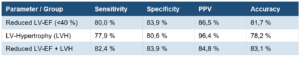
German Society for Cardiology - Cardiovascular Research e.V – 91st Annual German Cardiac Society Meeting 2025 – Evaluation of new vector electrocardiography algorithms for identifying patients with reduced left ventricular ejection fraction and left ventricular hypertrophy
![]()
Originally presented at the 91st annual German Cardiac Society meeting 2025.
Background:
Reduced left ventricular ejection fraction (LV-EF) and left ventricular hypertrophy (LVH) are associated with high mortality and morbidity. This study evaluated the diagnostic performance of a modified vectorcardiography (VCG) algorithm, a technique which records cardiac electrical activity in three dimensions, for detecting reduced LV-EF and LVH.
Methods:
This study included patients with reduced LV-EF (< 40%) and LVH (indexed LV mass > 55 g/m²) compared with controls without cardiac pathology. Cardiac magnetic resonance imaging (CMR) was performed and used as the reference standard for measuring LV-EF and LV mass. The modified VCG analyzed 533 VCG parameters per heartbeat to classify cardiac status. In total, 533 regular parameters and 50 Frenet-Serret parameters were analyzed, amounting to 583 parameters. Parameters were selected based on a low p-value from the Mann-Whitney-Wilcoxon test, indicating statistical significance. Bayes’ theorem was used to update prior probabilities and to derive posterior probabilities for reduced LV-EF and LVH when compared with controls.
Results:
A total of 280 patients were included. The group with reduced LV-EF (n=40) had a mean age of 56 ± 16 years and was predominantly male (78%), with a median LV-EF of 31.5% (23.4-36.3%). The LVH group (n=209) had a mean age of 60 ± 16 years (87% male) with an indexed LVM of 67g/m² (61-77 g/m²). Controls (n=31) had a mean age of 50 ± 16 years (61% male), normal LV-EF of 62 ± 5.6% and indexed LV mass of 40g/m² (35-47g/m²). In total 4 VCG parameters were used. A sensitivity of 80.0%, a specificity of 85.7% and an accuracy of 82.9% were achieved for the detection of reduced LV-EF. A Sensitivity of 74.5%, a specificity of 68.6% and an accuracy of 73.6% were reached for the detection of LVH. In with patients haing both reduced LV-EF and LVH (n=64), the VCG demonstrated a sensitivity of 79.3% and a specificity of 85.7% with an accuracy of 82.8%.
Conclusion:
The modified VCG algorithm showed notable diagnostic value for detecting reduced LV-EF and LVH. The VCG could serve as a fast, non-invasive, and cost-effective method to assist clinicians in identifying significant cardiac pathologies and guiding further diagnostic steps. Further optimization of the VCG algorithm is necessary to improve differentiation between healthy individuals and those with cardiac disease, enhancing its clinical use.
Estudios y literatura adicionales sobre nuestra tecnología.
The Poynting vector: power and energy in electromagnetic fields
Carpenter KH. The Poynting vector: power and energy in electromagnetic fields. Departement of Electrical and Computer Engineering. Kansas State University (2004)
Spatial vector electrocardiography; the clinical characteristics of S-T and T vectors
Grant RP, Estes EH Jr, Doyle JT. Spatial vector electrocardiography; the clinical characteristics of S-T and T vectors. Circulation. 1951 Feb;3(2):182-97.
Cardiogoniometry: a new noninvasive method for detection of ischemic heart disease
Saner H, Baur HR, Sanz E, Gurtner HP. Cardiogoniometry: a new noninvasive method for detection of ischemic heart disease. Clin Cardiol. 1983 May;6(5):207-10.
Kardiogoniometrie: eine elektrokardiografische, nichtinvasive und belastungsfreie Methode zur Erkennung der kardialen Ischämie
Sanz, Ee. Schüpbach, M. Kardiogoniometrie: eine elektrokardiografische, nichtinvasive und belastungsfreie Methode zur Erkennung der kardialen Ischämie. GMS Medizinische Informatik, Biometrie und Epidemiologie. 5 (2009).
Applicability of cardiogoniometry as a non-invasive screening tool for the detection of graft vasculopathy in heart transplant recipients
Spiliopoulos S, Hergesell V, Fischer D, Dapunt O, Krueger U, Koerfer R, Tenderich G. Applicability of cardiogoniometry as a non-invasive screening tool for the detection of graft vasculopathy in heart transplant recipients. Interact Cardiovasc Thorac Surg. 2016 Dec;23(6):976-978.
Computeranalyse des korrigierten, orthogonalen Kardiogramms
von Mengden, H.J., Brodda, K. Computeranalyse des korrigierten, orthogonalen Kardiogramms. Archiv für Kreislaufforschung 67, 123–141 (1972).
Assessment of the Spatial QRS-T Angle by Vectorcardiography: Current Data and Perspectives
Voulgari C, Tentolouris N. Assessment of the Spatial QRS-T Angle by Vectorcardiography: Current Data and Perspectives. Curr Cardiol Rev. 2009 Nov;5(4):251-62. doi: 10.2174/157340309789317850. Erratum in: Curr Cardiol Rev. 2010 Nov;6(4):373.
Improved evaluation of left ventricular hypertrophy using the spatial QRS-T angle by electrocardiography
Maanja, M., Schlegel, T.T., Kozor, R. et al. Improved evaluation of left ventricular hypertrophy using the spatial QRS-T angle by electrocardiography. Sci Rep 12, 15106 (2022).